
Neonatology · Chest wall · Practical review
A depressed sternum may be an incidental shape or compromise breathing: appearance, radiographic indices and clinical function must be interpreted together.
Key points. Pectus excavatum is a dorsal deviation of the sternum and costal cartilages that reduces chest depth. Some young animals have only a visible deformity; others develop rapid or laboured breathing, nursing or exercise intolerance, poor growth, respiratory disease or cardiac displacement. Radiographs confirm and quantify the deformity, but no single index decides whether surgery is required. CT better defines asymmetry, lung volume and complex surgical anatomy. In a symptomatic patient with a still-compliant chest, early correction can be very effective, but it requires an experienced team and close follow-up.
Information for breeders and owners. This is neither a splinting guide nor a surgical protocol: uncontrolled pressure or traction can injure the lungs, heart, major vessels and skin.
1. A sternal deformity, not merely a “flat chest”
Pectus excavatum is a dorsal displacement—towards the spine—of part of the sternum and adjacent costal cartilages. Local dorsoventral chest depth is reduced. The depression may be caudal or more extensive, central or lateralised, symmetrical or associated with sternal torsion. This geometry helps explain why two patients that look similar externally may have very different respiratory consequences.
The condition occurs in dogs and cats, although most contemporary clinical evidence concerns cats. It is usually recognised during the first weeks or months of life, but a moderate deformity may be found later. It should be distinguished from:
- a more diffuse chest flattening in a puppy or kitten lying continuously in sternal recumbency;
- swimmer puppy syndrome, primarily a locomotor disorder that may coexist with a thoracic deformity;
- pectus carinatum, in which the sternum protrudes outwards;
- an acquired deformity after trauma or metabolic bone disease.
Important distinction. “Congenital” means present during early development; it does not prove a heritable disorder. Describe the exact phenotype, look for associated anomalies and document the litter before drawing breeding conclusions.
2. From cosmetic shape to cardiopulmonary compromise
A shallow depression may remain clinically silent. When intrathoracic space is meaningfully reduced, lung expansion may be limited and the heart may be displaced or compressed. Signs depend on age, chest-wall rigidity, the location of the depression and concurrent disease.
| Observation | Possible meaning | Action |
|---|---|---|
| Palpable depression; bright patient; quiet breathing and normal growth | Mild or well-compensated form, although severity cannot be established by palpation alone. | Arrange an early assessment, weigh and photograph consistently; image as guided by the examination. |
| Tachypnoea, poor stamina, pauses during nursing or play | Restrictive ventilation, cardiac displacement/compression, pain or concurrent respiratory disease. | Same-day or prompt assessment depending on intensity. |
| Cough after nursing, regurgitation, nasal discharge, fever or depression | Aspiration pneumonia, gastrointestinal anomaly, infection or another complication—not necessarily the deformity alone. | Targeted evaluation and thoracic imaging without waiting for positional correction. |
| Marked dyspnoea, cyanosis, inability to nurse or collapse | Cardiopulmonary decompensation or a concurrent emergency. | Emergency oxygenation and stabilisation before corrective planning. |
In a 2026 retrospective series of 25 cats younger than 20 weeks treated with external coaptation, 19 presented with tachypnoea or exercise intolerance. This referral population does not represent every pectus case, but it confirms that functional signs matter at least as much as measurements. [7]
3. Examination links chest shape to patient function
Assessment starts while the patient is calm. The veterinarian observes respiratory rate and pattern, symmetry, mucosal colour, posture, ability to nurse or walk, then auscultates the lungs and heart. Palpation identifies compliance, the point of maximal depression, asymmetry, pain and any rib deformity.
The work-up extends beyond the sternum. Depending on presentation it includes:
- weight and growth trajectory, hydration, temperature and nutritional state;
- orthopaedic and neurological assessment when the limbs splay or the patient cannot stand;
- search for a murmur, arrhythmia, weak pulses, cyanosis or exercise intolerance;
- palate, swallowing and oesophageal assessment when regurgitation or aspiration is suspected;
- search for vertebral, rib, diaphragmatic or abdominal anomalies.
Short home videos of resting respiration, nursing and walking on a non-slip surface can help document change. They cannot reliably measure oxygen saturation or exclude pneumonia or heart disease.
4. Radiographs, indices and CT: measure without reducing the patient to a number
Thoracic radiography
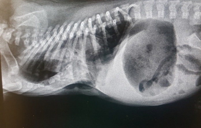
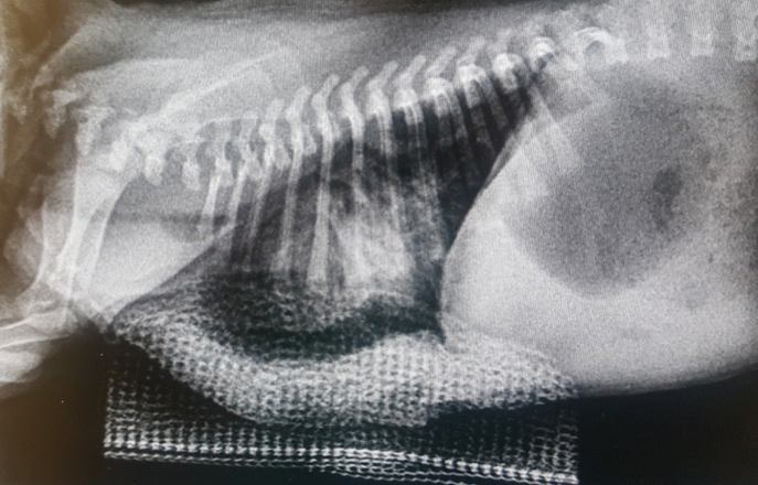
Good-quality orthogonal views confirm sternal deviation, locate its maximum and assess the lungs, cardiac silhouette, ribs and vertebrae. Reproducible positioning matters because rotation, poor inspiration and obliquity can alter measurements. In a dyspnoeic patient, safety takes priority over a perfect image.
Vertebral and frontosagittal indices
The vertebral index relates chest depth to vertebral-body height; the frontosagittal index relates chest width to depth. They may be measured at the point of maximal deformity and/or at a reference level, often T10, depending on the published method. A marked depression generally lowers the vertebral index and raises the frontosagittal index.
“Normal” ranges depend on species, chest conformation, breed, age, level and method; they must not be transferred mechanically. In a prospective cohort of ten kittens, radiographic indices approximated CT dimensions but correlated poorly with clinical severity. In 12 brachycephalic dogs, location and symmetry also contributed to clinical presentation. [3] [4]
CT and complementary imaging
CT provides a three-dimensional view, measures asymmetry and torsion, defines relationships between the sternum, heart, major vessels and lungs, and can estimate chest and lung volumes. It is especially valuable before complex, internal, thoracoscopic or patient-specific correction. A CT study of 14 cats highlighted that the deformity may be more complex than a single reduced diameter. [5]
The expected benefit must be balanced against transport, restraint, sedation or anaesthesia in a very young respiratory patient. Echocardiography is indicated when a murmur, congenital heart defect, substantial cardiac displacement/compression or perioperative functional concern is present. Thoracic ultrasound can answer selected pulmonary questions but does not describe the entire chest-wall deformity.
| Test | Useful information | Main limitations |
|---|---|---|
| Radiographs | Confirmation, level, indices, lungs, heart, ribs and vertebrae; accessible follow-up. | Two-dimensional projection; position and inspiration dependent; indices correlate imperfectly with signs. |
| CT | Asymmetry, torsion, volumes and organ/vessel relationships; 3D planning. | Availability, cost, radiation and usually sedation/anaesthesia; unnecessary for every mild case. |
| Echocardiography | Cardiac anatomy and function, effect of displacement, associated heart defects. | Windows may be altered; does not characterise the entire skeletal deformity. |
| Standardised follow-up imaging | Objective comparison during growth and correction. | Requires consistent views and method; never replaces clinical change. |
Avoid the shortcut. Do not call a case “mild” solely because the patient is quiet in clinic, or “severe” solely because one number crosses a published threshold. Home breathing, feeding, growth, asymmetry and concurrent disease must guide the decision.
5. A targeted work-up: investigate what can change management
Testing is led by clinical questions, not an automatic panel. Haematology, biochemistry and glucose can be useful before anaesthesia or in a weak neonate, but they do not measure mechanical severity. Pulse oximetry can document oxygenation, although motion, perfusion and patient size affect a single value. Blood gases, culture and other tests are reserved for situations in which they answer a defined question.
| Question | Conditions to consider | Possible targeted tests |
|---|---|---|
| Is the chest shape the only respiratory problem? | Pneumonia, oedema, atelectasis, pulmonary malformation, infection, upper-airway obstruction. | Auscultation, radiographs/CT, oxygenation and indicated analyses or sampling. |
| Is the heart merely displaced or also abnormal? | Murmur, congenital defect, compression, arrhythmia. | Echocardiography, ECG and blood-pressure assessment as indicated. |
| Is a locomotor disorder present? | Swimmer puppy syndrome, luxation, pain, vertebral anomaly, neurological/neuromuscular disease. | Orthopaedic and neurological examination, targeted radiographs and specialist assessment. |
| Could another malformation explain the presentation? | Pectus carinatum, diaphragmatic or peritoneopericardial hernia, rib/vertebral anomaly, trauma. | Appropriate radiography, ultrasound or CT. |
A diagnosis of pectus does not exclude a second disease. Conversely, treating pneumonia or improving locomotor support may substantially change the clinical picture and clarify what remains attributable to the chest wall.
6. Monitor or correct: combine function, anatomy and age
Structured monitoring can be reasonable for a shallow, stable and genuinely asymptomatic deformity after a baseline examination, with a growth record and reassessment plan. “Waiting” does not mean ignoring: the chest wall becomes less compliant with age, while respiratory deterioration may increase anaesthetic risk.
Correction is more strongly considered when several findings converge:
- dyspnoea, persistent tachypnoea or nursing/exercise intolerance;
- poor growth or respiratory episodes plausibly related to the deformity;
- moderate to severe, central, progressive or markedly asymmetric anatomy;
- organ displacement/compression or reduced lung volume on imaging;
- a still-mobile sternum that offers a less invasive corrective window;
- persistent signs after treatment of concurrent disease and optimisation of support.
There is no single ideal age for every patient. Chest compliance, safe anaesthetic size, intensity and progression of signs, and the planned technique must all be balanced. Dr Xavier Lévy’s paediatric surgery material stresses the value of early assessment before the deformity becomes rigid, while still requiring an appropriate cardiopulmonary work-up.
How specialist input can save time. The aim is not to order every test. It is to select imaging that answers the actual question, determine whether the deformity explains the signs, and choose the right timing and technique.
7. From supportive care to corrective surgery
Medical and environmental support
Support addresses consequences and concurrent disorders: oxygen and stabilisation for distress, evidence-led treatment of infection, safe nutritional support, aspiration prevention, a non-slip surface and physiotherapy when swimmer syndrome coexists. These measures do not necessarily realign a structurally displaced sternum.
Monitoring a mild form
Monitoring combines serial weight, sleeping respiratory rate, nursing and activity tolerance, physical examination and follow-up imaging when shape changes. Families need written triggers for seeking care before the planned revisit.
External coaptation
In a young patient with a compliant chest, sutures placed around sternal segments can provide outward traction against a moulded external splint. Casting or 3D printing may distribute forces and accommodate asymmetry. The principle sounds simple, but sutures pass immediately adjacent to lungs, heart and vessels; this is never a home-made restraint.
In the August 2026 retrospective series of 25 young cats, 24 survived, radiographic conformation improved and signs had resolved at recheck. Pneumothorax, suture breakage, dermatitis, residual defect and one death nevertheless occurred. The study had no untreated comparison group and came from a referral centre. [7]
Thoracoscopic visualisation
An intrathoracic camera can show needle passage and the deep structures during suture placement. Six kittens treated at two centres in 2026 all recovered from anaesthesia, but one experienced respiratory arrest 36 hours later for an undetermined reason. A 2026 three-cat series and a 2025 single case support feasibility, not proven superiority. [8] [9]
Internal or combined correction
A plate secured to the sternum, an internal construct or an open/assisted technique may be chosen when the sternum is rigid, the deformity is severe or simple external traction is unsuitable. In a 2026 series of six cats and one dog, imaging showed immediate increases in chest and lung volumes; one pneumothorax, one sternebral fracture and one clinically silent cerclage breakage occurred. Two cats later had reduced lung volume with new pulmonary disease. Seven patients cannot establish a universal standard. [10]
Progressive correction with a patient-specific external splint has been described in a severe single case to reduce the risk of abrupt re-expansion. A historical report of fatal re-expansion pulmonary oedema after rapid correction supports individualising both correction rate and monitoring. [6] [13]
| Option | Possible setting | Evidence and limitations |
|---|---|---|
| Structured monitoring | Shallow, stable and asymptomatic after assessment. | Clinical reasoning; natural-history data remain sparse, so follow-up is essential. |
| Support/physiotherapy | Concurrent disease or swimmer syndrome; preoperative stabilisation. | Useful for associated problems, but may not correct the sternum. |
| External splint | Young, compliant chest with a clinically important deformity. | Largest current series: 25 retrospective cats; favourable results with real complications; limited canine data. |
| Thoracoscopic assistance | Direct visualisation of suture passage and intrathoracic structures. | Case reports and small series; feasible, not robustly compared. |
| Internal plate/combined technique | Rigid, severe, asymmetric or unsuitable for simple external traction. | Cases and a seven-animal series; specialist surgery, implant risks and limited long-term evidence. |
8. Follow-up is part of treatment
Monitoring starts during anaesthesia and continues throughout growth under correction. Potential complications include:
- pneumothorax, haemorrhage or injury to lung, heart, diaphragm or vessels during suture passage;
- postoperative hypoventilation, atelectasis, pain or overly rapid pulmonary re-expansion;
- skin pressure, dermatitis, wounds, infection, moisture or splint displacement;
- breakage, loosening, migration or exposure of the construct;
- insufficient, excessive or asymmetric correction;
- recurrence after removal, particularly while growth and contributing factors continue.
At home, caregivers need written monitoring criteria: sleeping respiration, mucosal colour, appetite, comfort, weight, odour or discharge, swelling, visible skin, device position and activity. Any sudden change warrants immediate contact. A rapidly growing patient can become constricted even when the splint does not look displaced.
Clinical and radiographic rechecks assess both shape and function. Removing or adjusting a device by calendar alone, without re-examining the patient, risks inadequate correction or avoidable complications.
9. Prognosis: often favourable in selected young patients, but evidence remains limited
Published cases and recent series frequently report substantial improvement in breathing, activity tolerance and conformation after correction. The prospective ten-kitten cohort described excellent medium-term outcomes in six of seven severely affected kittens that underwent splinting, while the 2026 series strengthen that favourable signal. [3] [7] [10]
Limitations must be clear: small samples, predominantly feline evidence, referral selection, heterogeneous techniques, no randomised comparison and often short follow-up. A good radiograph does not guarantee normal pulmonary function, and a new respiratory disease can alter outcome. Prognosis depends on timing, chest compliance, functional severity, concurrent anomalies and access to close follow-up.
An unoperated mild form may remain compatible with normal life, but natural-history evidence is sparse. Monitoring is therefore an active strategy, not proof that the deformity is harmless.
10. Probably multifactorial: reason at litter level
The exact cause remains uncertain. Abnormal sternal/cartilage development, mechanical forces, rapid growth and familial susceptibility have been proposed. An increased incidence of thoracic-wall deformities has been reported among related Bengal kittens, and two littermate dogs have been described. These observations support a possible predisposition but neither prove simple inheritance nor provide a validated genetic test. [11] [12]
For breeding records, retain:
- photographs, imaging, diagnosis and outcome of the affected animal;
- the identity and status of every littermate, including neonatal deaths;
- both pedigrees, litter size, delivery conditions and nesting surface;
- other malformations, swimmer syndrome or respiratory signs.
Recurrence in the same family warrants pedigree review and a reproductive consultation before repeating the mating. Without a causal variant or robust breed-specific data, no universal breeding rule is defensible; severity, recurrence, relatives and genetic diversity all matter.
Related topic: swimmer puppy syndrome — a separate article currently being updated.
11. Practical questions
Does every sternal depression require surgery?
Can I massage or press the sternum into place?
Do radiographic indices show whether the patient is suffering?
Does every case need CT?
Is pectus excavatum the same as swimmer puppy syndrome?
How long is a splint kept in place?
Should the animal be removed from breeding?
Specialist assessment
When breathing or chest shape raises concern
Dr Xavier Lévy can coordinate examination, imaging and strategy with your primary-care veterinarian at CRECS near Toulouse or Bordeaux Sud, and provide FR/EN case-document review remotely where regulations allow.
12. References and strength of evidence
Dr Xavier Lévy’s personal sources consulted for this review: Surgery EVSSAR Xavier Levy 2026 (16 March 2026) and Pediatric surgery X Levy (4 September 2025), sections on radiographic assessment and external/internal correction; personal copy of Crigel & Moissonnier 2005. Surgical details and drug doses were not reproduced. A human surgery paper located in the same folder was inspected but not used as veterinary evidence.
1. Fossum TW, Boudrieau RJ, Hobson HP, et al. Surgical correction of pectus excavatum, using external splintage in two dogs and a cat. J Am Vet Med Assoc. 1989;195:91–97. Foundational three-case series. PMID 2759902.
2. Crigel MH, Moissonnier P. Pectus excavatum surgically repaired using sternum realignment and splint techniques in a young cat. J Small Anim Pract. 2005;46:352–356. Single case. DOI 10.1111/j.1748-5827.2005.tb00332.x.
3. Charlesworth TM, Schwarz T, Sturgess CP. Pectus excavatum: computed tomography and medium-term surgical outcome in a prospective cohort of 10 kittens. J Feline Med Surg. 2016;18:613–619. DOI 10.1177/1098612X15591234.
4. Hassan EA, Hassan MH, Torad FA. Correlation between clinical severity and type and degree of pectus excavatum in twelve brachycephalic dogs. J Vet Med Sci. 2018;80:766–771. DOI 10.1292/jvms.17-0518; PMID 29593167.
5. Komsta R, Łojszczyk A, Dębiak P, et al. Computed tomographic evaluation of pectus excavatum in 14 cats. PLoS One. 2022;17:e0262866. Descriptive CT study. DOI 10.1371/journal.pone.0262866; PMID 35061860.
6. Zanfabro M, et al. Use of a customized 3D-printed external splint for correction of severe pectus excavatum in a 3-month-old kitten. JFMS Open Rep. 2022;8. Single case with progressive correction. PMID 35342728.
7. Heidgerd KM, Casale S, Van Hatten R. Outcome and complications associated with correction of pectus excavatum using external coaptation in cats less than 20 weeks of age: 25 cases (2010–2024). Vet Surg. Online 13 August 2026. Retrospective study. DOI 10.1111/vsu.70142; PMID 42591014.
8. Jones G, et al. Experience with video-assisted thoracoscopic placement of pectus excavatum splints in kittens: six cases. J Small Anim Pract. 2026;67:830–835. Small uncontrolled series. DOI 10.1111/jsap.70142; PMID 42107986.
9. Charlesworth T. Cannula-assisted sternal displacement for video-assisted thoracoscopic surgery correction of pectus excavatum in three cats. JFMS Open Rep. 2026. Three cases. DOI 10.1177/20551169261440980.
10. Torres Villegas M, et al. Internal plate fixation successfully corrects pectus excavatum with minimal complications in 6 cats and 1 dog. J Am Vet Med Assoc. 2026. Seven animals with imaging follow-up. DOI 10.2460/javma.25.10.0645; PMID 41564546.
11. Charlesworth TM, Sturgess CP. Increased incidence of thoracic wall deformities in related Bengal cats. J Feline Med Surg. 2012;14:365–368. Familial association, no causal variant identified. DOI 10.1177/1098612X12437351.
12. Rahal SC, Morishin Filho MM, Hatschbach E, et al. Pectus excavatum in two littermate dogs. Can Vet J. 2008;49:880–884. Two related cases, limited evidence. PMID 19043484.
13. Fossum TW, et al. Fatal reexpansion pulmonary edema in a kitten following surgical correction of pectus excavatum. J Am Anim Hosp Assoc. 1995;31:133–136. Historical safety signal from a single case. PMID 7773758.
Critical interpretation. Most reports are single cases or small series and predominantly feline. The 2026 series of 25 cats is the largest identified for external coaptation, but it remains retrospective. Newer approaches are promising; current evidence cannot establish one superior technique for every patient.